

Address: 2100/2, Trichy Road,
Rajalakshmi Mills Stop, Singanallur,
Coimbatore, Tamil Nadu 641005
Surgical Management – A Rare Case Series
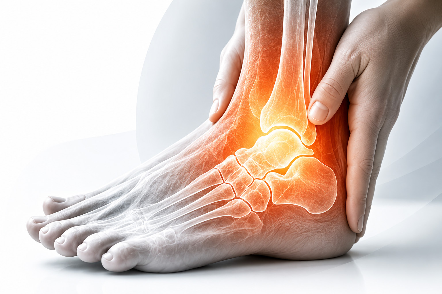
Ankle and subtalar arthritis – a rare but potentially disabling condition.
Ankle and subtalar arthritis are rare, but often disabling conditions. When conservative treatment fails, surgical intervention can restore stability, relieve pain, and improve quality of life.
This case series presents two complex patients who underwent ankle–subtalar fusion using a retrograde intramedullary nail technique, followed by structured postoperative rehabilitation.
A 60-year-old female presented with severe limping and walker dependency for 18 months following a road traffic accident. Initial X-rays revealed an ankle fracture, and surgery was advised.
However, she declined surgery and underwent native oil bandaging for over six months. Despite prolonged conservative treatment, she continued to experience severe pain and difficulty walking.
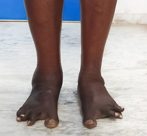
Case 1 – Clinical examination showing deformity, swelling and lower-limb changes.
Clinical examination revealed deformity and swelling around the ankle, severe ankle tenderness, severe subtalar joint tenderness and approximately 90% restriction of ankle and subtalar movements.
There was significant swelling, with no midfoot or distal tibia tenderness. Hyperpigmented patches were also present over both distal legs due to prolonged native medication use.
X-ray ankle AP and lateral views showed a malunited bimalleolar fracture ankle with ankle subluxation and reduced ankle and subtalar joint space.
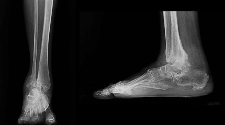
Case 1 – Preoperative X-ray showing the malunited bimalleolar fracture, ankle subluxation and reduced joint spaces.
The patient underwent ankle–subtalar fusion using a retrograde intramedullary nail technique.
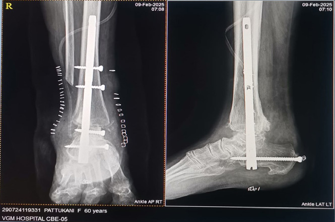
Case 1 – Surgical fixation following ankle–subtalar fusion.
The procedure presented several technical challenges, including difficult reduction due to malunion and chronic joint dislocation.
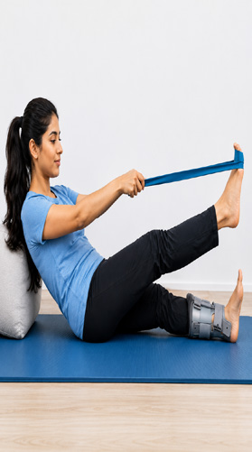
Structured postoperative rehabilitation shown in the supplied case material.
Three months later, the patient was walking without a limp, with full weight bearing. The supplied case presentation records continued walking without a limp for the following two years.

Case 1 – Reported recovery and functional walking outcome.
A 65-year-old female, a known case of rheumatoid arthritis, presented with progressively worsening ankle pain and limping for the past nine months.
She had previously undergone bilateral Total Knee Replacement (TKR) under the orthopaedic team in 2016 and remained pain-free following the procedure.
She was on Disease-Modifying Anti-Rheumatic Drugs (DMARDs), which effectively controlled her generalized body pain and polyarthralgia. However, her ankle pain progressively worsened, significantly affecting her mobility and quality of life.
Radiographic evaluation revealed severe osteoporosis, advanced arthritis involving both the ankle and subtalar joints, and severe arthritis with ankylosis of the midfoot tarsal bones.
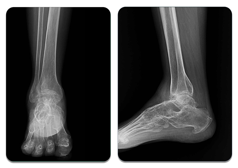
Case 2 – Radiographic evaluation showing advanced ankle and subtalar arthritis.
The patient underwent ankle–subtalar fusion using retrograde intramedullary nailing.
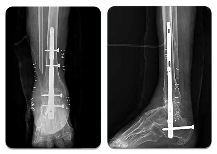
Case 2 – Surgical fixation following ankle–subtalar fusion using retrograde intramedullary nailing.
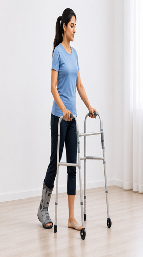
The supplied case presentation illustrates rehabilitation through staged strengthening, gait training, balance, cycling and functional activities.
Weeks 0–2
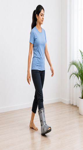
Weeks 2–6
Weeks 6–12
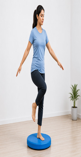
Functional Training
At the three-month follow-up, the patient achieved pain-free full weight bearing without a limp.
She continued to walk independently with excellent functional outcomes for 18 months following surgery.
Advanced ankle and subtalar arthritis can severely impair mobility. Retrograde nail ankle and subtalar fusion provides stable and durable fixation in appropriately selected complex cases.
Structured rehabilitation is essential for optimal recovery, while timely surgical intervention can help restore pain-free walking and improve quality of life.
Combined ankle and subtalar arthritis is a rare condition commonly associated with rheumatoid arthritis, post-traumatic injury or advanced degeneration.
Early cases may respond to medication, physiotherapy, bracing and activity modification. In advanced disease, ankle–subtalar fusion provides a surgical solution for pain relief, stability and restored mobility.
The two cases in this series demonstrate encouraging functional outcomes following ankle–subtalar fusion using retrograde intramedullary nailing, supported by structured postoperative rehabilitation.
Case 1: Walking without a limp with full weight bearing at three months, with continued walking without a limp documented for two years.
Case 2: Pain-free full weight bearing without a limp at three months, with independent walking and excellent functional outcomes documented for 18 months.
This article is based on the supplied VGM Hospital case-series material. Treatment decisions should be made after individual clinical and radiographic assessment by a qualified orthopaedic specialist.
— Dr Madura Prasad Suman
VGM Hospital, Trichy Road, Coimbatore
Call us Anytime at: +91 9843079555
Land Line No: 0422-2572573
Fax: 0422-2572207
Email: contact@vgmorthocentre.com
Hospital Timing: 24 x 7
OP Appointment Timing:
Mon-Fri: 9:00am-8:00pm
Legal Policy